
Detailed EBSD Study of a Bivalve Shell
An in-depth look at the structures of a mussel shell, characterised using Symmetry. Both calcite and nanostructured aragonite nacre are measured with an unprecedented level of detail and speed.
The improved sensitivity of the latest generation of fibre-optically coupled CMOS-based Electron Backscatter Diffraction (EBSD) detectors has made the analysis of biomaterials using EBSD far more routine. Many biomaterials, such as shell structures, are relatively sensitive to damage by the electron beam yet have small scale structures that require high resolution analyses. High speed, high resolution EBSD maps enable these complex structures to be characterised effectively, in turn improving our understanding of their formation and physical properties.
EBSD has been applied widely to a range of biomaterials, including for:
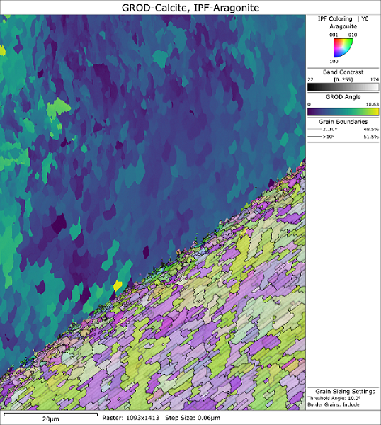
The image on the right-hand side shows an EBSD-derived map of the calcite (top) – aragonite (bottom) interface in a common mussel (Mytilus edulis) shell. Calcite is coloured using a grain relative orientation colouring scheme, with aragonite coloured using a standard inverse pole figure colouring scheme.
An in-depth look at the structures of a mussel shell, characterised using Symmetry. Both calcite and nanostructured aragonite nacre are measured with an unprecedented level of detail and speed.
© Oxford Instruments 2026